X-ray Imaging
X-ray Imaging
Fluoroscopic X-ray
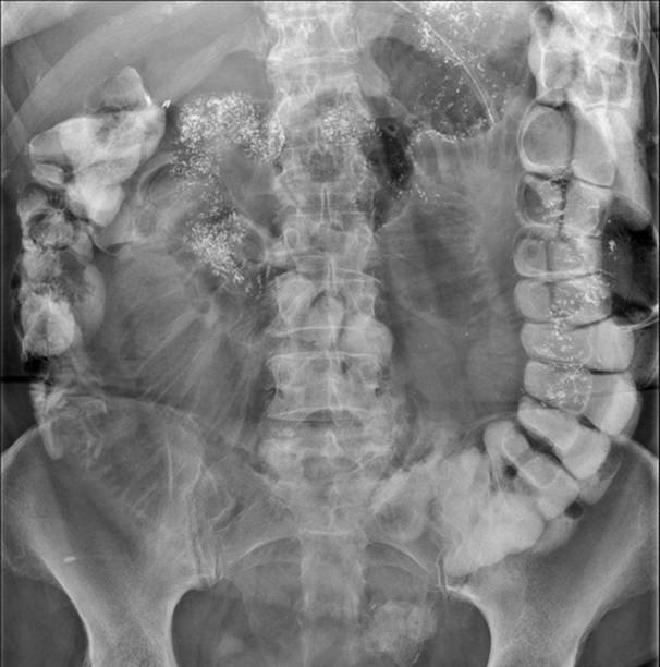
X-ray imaging is a diagnostic technique that uses a small dose of ionizing radiation to create pictures of the inside of the body. X-rays can be used for detecting broken bones or fractures, checking for lung infections, part of pre-op screening, identifying dental problems, or for diagnostic joint or spine issues.
Additionally, x-rays are used in fluoroscopic procedures. Instead of just taking a single snapshot (like a regular x-ray), fluoroscopy lets doctors watch how organs, tissues, or instruments move and function inside of the body during a procedure. These procedures include but are not limited to modified barium swallows, upper GIs, joint injections, myelograms, lumbar punctures, hysterosalpingograms and more.
In short, x-rays are mainly used to see inside the body without surgery, making them a powerful tool for diagnosis and treatment.
Considerations to take into account:
Preparation Instructions
Your preparation depends on the type of exam you have done.
- General x-rays do not typically require any preparation prior to the exam.
- You may be asked to change for your exam to remove any items or clothing that may interfere with your exam.
- Fluoroscopy exams vary, as will the preparation ahead of time depending on the exam your doctor orders. Your healthcare provider will order your exam, provide a brief description of prep for the procedure, and you will receive full directions for preparations at the time of scheduling.
Important Reminders
- Consult your doctor: Always discuss any concerns about the risks or suitability of a x-ray with your doctor.
- Provide medical history: Be sure your doctor is aware of all your medical conditions, medications, and allergies.
- Follow instructions: Adhere to any instructions given by your healthcare provider regarding attire, diet, medication and other preparations for your x-ray.