Lung Cancer Treatments and Procedures

The TriHealth Cancer & Blood Institute offers several treatments for lung cancer, including Chemotherapy and Radiation as well as surgery to remove all or part of a tumor.
Surgery
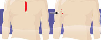
Surgery for lung cancer is known as a lobectomy. Similar to “open heart” surgery, it traditionally involves sternotomy – cutting through the breastbone and opening the ribs. This can cause significant trauma, prolong healing time and increase the risk for serious complications and even mortality.
Surgery depends upon your individual situation. This is a decision made in collaboration with the tumor board and your health care team.
Lung Cancer Robotic-Assisted Lobectomy is Less Invasive
Robotic-assisted lobectomy is an alternative to traditional open surgery. When performed robotically with the da Vinci Surgical System, lobectomy is done with unparalleled precision and control through a few small incisions along the side of the chest.
Benefits of Robotic-Assisted Lobectomy for Lung Cancer
In addition to avoiding the pain and trauma of sternotomy and rib spreading, this may provide patient benefits such as:
- Less risk of infection
- Less blood loss and need for blood transfusions
- Shorter hospital stay
- Significantly less pain and scarring
- Faster recovery
- Quicker return to normal activities
As with any surgery, these benefits cannot be guaranteed, as surgery is both patient- and procedure-specific. While robotic-assisted lobectomy is considered safe and effective, it may not be appropriate for every individual. Always ask your doctor about all treatment options, as well as their risks and benefits.
Other Procedures
Other procedures used during lung cancer diagnosis and treatment can include CT and PET scans as well as Chest X-Rays.
Chest X-Ray
A Chest X-ray is a painless, noninvasive test that creates pictures of the structures inside your chest, such as your heart, lungs, and blood vessels. "Noninvasive" means that no surgery is done and no instruments are inserted into your body.
This test is done to find the cause of symptoms such as shortness of breath, chest pain, chronic cough (a cough that lasts a long time), and fever.
X-rays are electromagnetic waves. They use ionizing radiation to create pictures of the inside of your body.
A Chest X-ray takes pictures of the inside of your chest. The different tissues in your chest absorb different amounts of radiation.
Your ribs and spine are bony and absorb radiation well. They normally appear light on a Chest X-ray. Your lungs, which are filled with air, normally appear dark. A disease in the chest that changes how radiation is absorbed also will appear on a Chest X-ray
When is a Biopsy introduced?
Biopsy can come at any time during this process. If/when a mass is discovered it is quickly tested to determine the course and treatment of your illness. As your treatment progresses, your nurse remains in close contact. The nursing team is able to help you to navigate the system and to make choices that work for you. Importantly, most patients see multiple experts and learn about options.
Precision Oncology
Delivering personalized treatment based on your unique cancer genomics and biomarkers.
If you have a cancer diagnosis, you are seeking world-class care close to home. Because every cancer is unique, our innovative medical oncologists utilize state-of-the-art testing to select the right treatment plan for you. Learn more.
Illustrations courtesy of Intuitive Surgical, Inc.